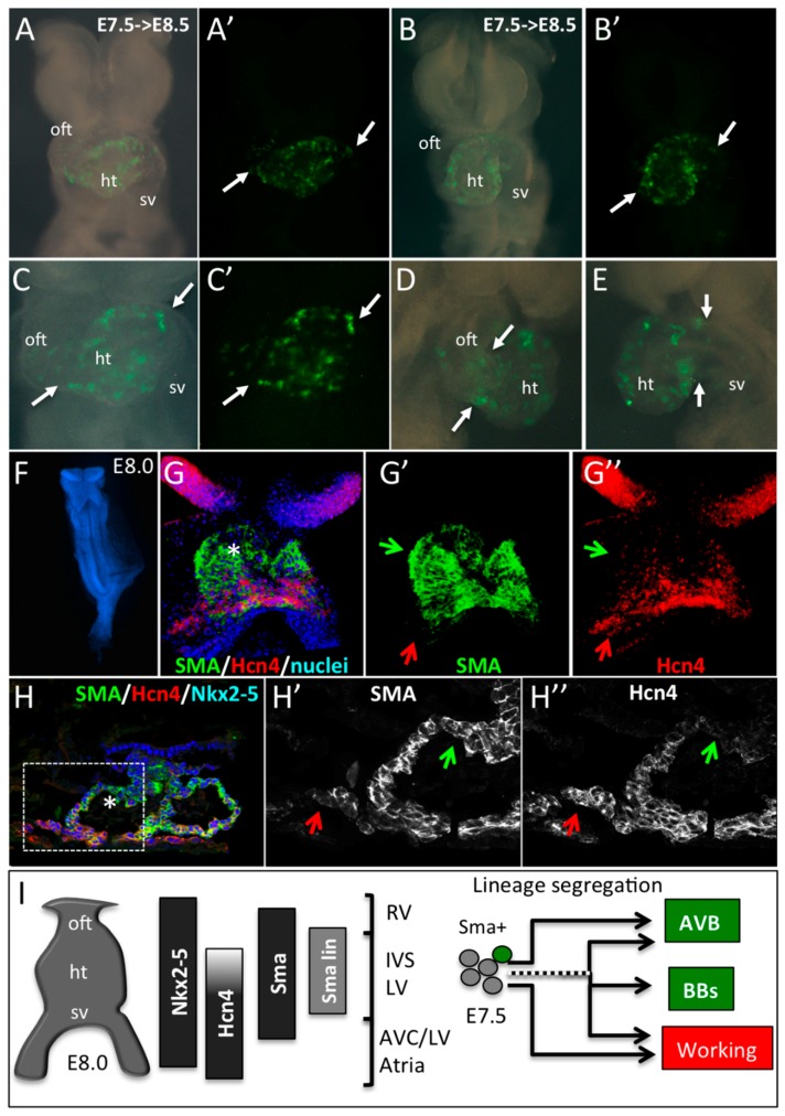
Figure 4.
Early SMA+ cardiomyocytes form the primary heart tube. (A–E) Whole-mount bright field or YFP fluorescence (A’–C’) of E8.5 SmaCre/+::R26RYFP/+ embryonic hearts induced by tamoxifen injection at E7.5. Ventral (A–C), right lateral (D) or left lateral (E) views show the presence of YFP+ cells in the heart tube only (ht). Arrows indicate the limit of YFP expression in the heart tube and regions with no labeling such as the outflow tract (oft) and sinus venosus (sv). (F–H) Immunofluorescence on whole-mount (G,G’,G’’) and sections (H,H’,H’’) of E8.0 embryos reveals a co-expression of SMA and Hcn4 in the lower part and a SMA+/Hcn4- expression domain in the upper part of the heart tube (star in G,H). An Hcn4 expression domain is observed at the venous pole (red arrows). Green arrows indicate the presumptive position of the early SMA+ cardiomyocytes in E8.0 heart tube. (I) Schematic representation of the expression domains of Nkx2-5, Hcn4, SMA and E7.5 SMA+ lineage (SMA lin) in the linear heart tube of an E8.0 mouse embryo. The compartments of the heart derived from different subdomains of the early heart tube are shown: RV, Right ventricle; IVS, Interventricular septum; LV, Left ventricle; AVC, Atrioventricular canal, and Atria. Our data suggest that precursors of the central VCS are included in the SMA+/Hcn4- region of the heart tube. While some early SMA+ cardiomycytes have already segregated to the conduction lineage to form the AVB, others, including cells giving rise to the bundle branches (BB), segregate later in embryonic development.

