Fig. 2.
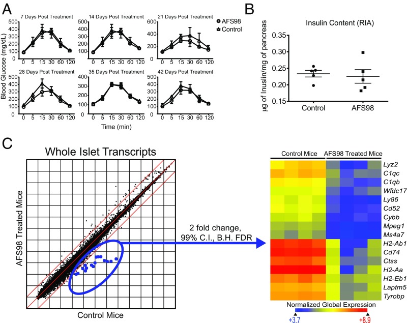
Islet function after depletion of macrophages by AFS98. (A) B6 mice were given 2.0 mg of AFS98 i.p. at 6–8 wk of age. Glucose tolerance assays were then performed on AFS98-treated and untreated mice. After the indicated number of days, the mice were fasted for 12 h and then injected with 2.0 g/kg glucose i.p. Blood glucose (mg/dL) was measured at the indicated time points. Results are pooled from two independent experiments (n = 2–3 mice per group). (B) Nine-week-old B6 females were left untreated or administered 2.0 mg of AFS98 i.p. Four days later, the mice were placed on a 20% sucrose diet for an additional 7 d, then returned to a normal diet for 2 d. The mice were then killed, and the insulin content of their islets was measured (n = 5 mice per group). (C) Three-week-old C57BL/6 mice were left untreated or administered 2.0 mg of AFS98 i.p. At 6 wk of age, their islets were isolated, total RNA was extracted, and transcripts were analyzed by microarray. Scatter plot shows the log2 mean expression values for four control and experimental mice. The dots highlighted in blue represent genes differentially expressed between treated and control mice at 99% confidence using moderated t test with Benjamini–Hochberg false discovery rate analysis. The selected genes are plotted in the heat map using Euclidean distance and normalized global expression as indicated.

