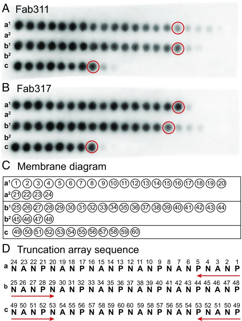
Fig. 2.
Epitope mapping using truncation peptide arrays. (A and B) The PepSpot membrane is shown for Fab311 (A) and Fab317 (B) and consists of five rows of the spotted peptides (a1, a2, b1, b2, and c). Dark spots indicate strong Fab binding. (C and D) Schematic of the location of the peptide spots on the membrane. The numbers within the circles refer to the numbers in the truncation array sequence (D). Rows a1 and a2 correspond to a truncation array starting from the C terminus of the (NANP)6 peptide, rows b1 and b2 are truncations from the N terminus, and row c represents truncations from both the N terminus and C terminus simultaneously. The peptides that appear to have the minimal number of repeats for strong Fab binding are circled in red in A and B.

