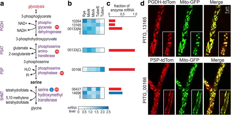
Fig. 7.

Serine biosynthesis in P. infestans. a, Enzymes for forming serine. Mitochondrial and cytosolic enzymes are marked by red and blue circlar symbols, respectively. b, mRNA levels in different tissues, as described in Fig. 1. For enzyme activities produced by more than one gene, the sum of CPM values in each tissue type is represented by the row labeled Σ. c, Contribution of each gene to the total transcript pool for each enzyme. d, Localization of enzymes. Top row, transformant co-expressing GFP-tagged mitochondrial marker and PGDH from PITG_00132 fused to tdTomato. Bottom, transformant co-expressing GFP-tagged mitochondrial marker and PSP from PITG_13749 fused to tdTomato. The smaller insets indicate alternative morphologies of mitochondria in P. infestans. The organelles are typically elongated in actively growing hyphae and rounder in dormant or slowly-growing cultures
