Figure Fig. 1.

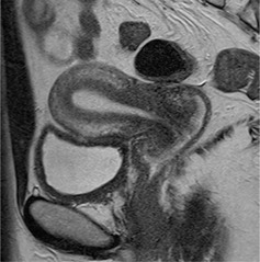
Representative case of a 34‐year‐old woman with a normal uterus. On T2‐weighted images, the uterus shows three distinct layers: a high signal endometrium, a low signal junctional zone, and an intermediate‐signal outer myometrium

Representative case of a 34‐year‐old woman with a normal uterus. On T2‐weighted images, the uterus shows three distinct layers: a high signal endometrium, a low signal junctional zone, and an intermediate‐signal outer myometrium