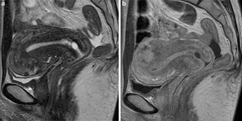
Figure Fig. 3.

Normal uterus of a 48‐year‐old woman. a On T2‐WI, the zonal anatomy of the uterine body can be recognized, but small and irregular low signal areas are observed within the anterior wall, indicating sporadic uterine contractions. b Ten minutes later (represented in a) the uterus appearance, imaged in contrast‐enhanced T1‐WI, shows differences from that seen on T2‐WI. A focal myometrial lesion with irregular enhancement has emerged in the posterior wall. In contrast, the thickness of the anterior wall looks reduced. These findings mean that the sporadic contraction of the anterior wall shown in a has disappeared and emerged in the posterior wall
