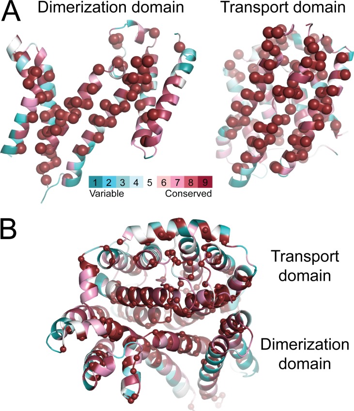
Figure 5.
Conservation of the residues at the interface between the transport and dimerization domains of AE1. (A and B) The x-ray structure (A), showing each domain as viewed from the other domain, and the repeat-swapped model (B), viewed from the intracellular side of the membrane. Residues are colored according to conservation from blue to burgundy, and those with the maximum conservation level are also indicated by spheres. Conservation was computed with the ConSurf webserver (http://consurf.tau.ac.il) using a PSI-BLAST search on January 4, 2016.

