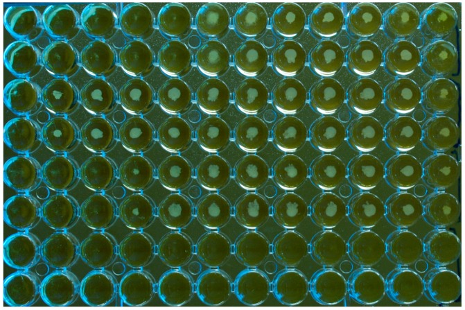
Figure 3.
Microplate (U-bottom wells) for susceptibility testing of a M. pachydermatis isolate against three antifungal drugs tested in duplicate, MCZ (rows 1 and 2); TBD (rows 3 and 4); CTZ (rows 5 and 6). Growth medium: Christensen Broth with Tween 40 and 80 as a lipid source. The last column on the right comprises the drug-free growth controls. The last column on the left is the negative control (no yeast inoculum; no drug) and the two bottom rows are also inoculum free. The rows of wells contain doubling dilutions of the drugs from 16 to 0.03 µg/mL with the highest concentration to the left of the plate. For this experiment, the MICs were 2, >16, and 4 µg/mL, respectively, for MCZ, TBD, and CTZ. The plate was not agitated prior to reading. It can be seen how the yeast isolate formed button-like deposits in the wells.

