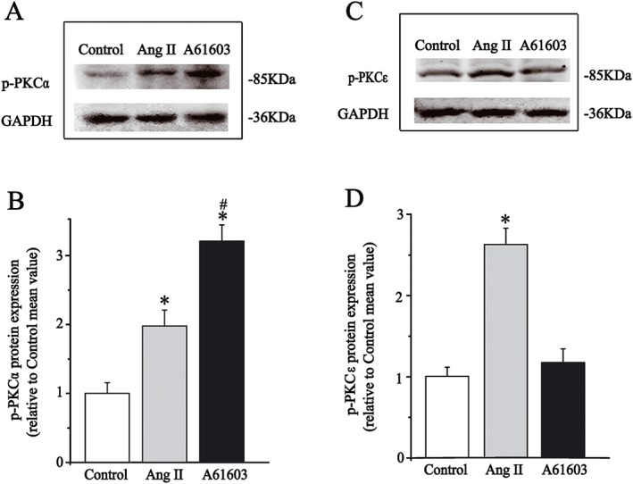
Figure 4.

Effects of A61603 and Ang II on the expression of phosphorylated PKC isoforms in isolated guinea pig ventricular cardiomyocytes. (A, C) Representative immunoblots for phosphorylated PKCα (left) and PKCε (right) along with internal standard GAPDH after application of A61603 or Ang II for 10 min. (B, D) Summary data for expression levels of phosphorylated PKC isoforms that were presented as fold change compared to control group mean, and the quantification of the band intensities were normalized with GAPDH. All data were obtained from five guinea pig hearts, *P < 0.05, significantly different from control; # P < 0.05, significantly different from Ang II.
