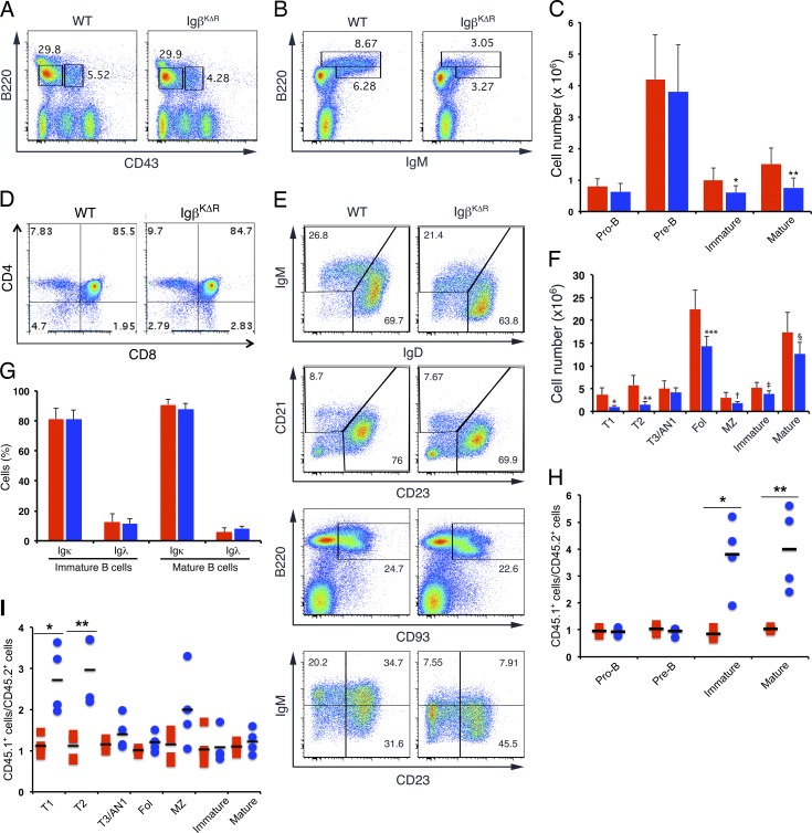
Figure 1.
Selective defect in late B cell development in IgβKΔR mice. (A–C) BM cells from WT and IgβKΔR mice were isolated, stained with antibodies specific for B220, CD43, and IgM, and analyzed by flow cytometry (A and B). Total numbers of each population (C) are provided for WT (red) and IgβKΔR (blue) mice; error bars indicate mean ± SD. *, P = 0.0167; **, P = 0.0022 (n = 3). (D) Thymic CD3+ lymphocytes were analyzed by flow cytometry for CD4 and CD8 expression (n = 3 mice). (E and F) Splenic B cells from the indicated mice were stained with antibodies specific for B220, IgM, IgD, CD21, CD23, and CD93 and then analyzed by flow cytometry. Representative flow cytometric plots are shown in E, and total numbers of each population from WT (red) and IgβKΔR (blue) are shown in F. *, P = 0.0046; **, P = 0.0021; ***, P = 0.0044; †, P = 0.0009; ‡, P = 0.0012; §, P = 0.0033 (n = 4 mice per condition). (G) Flow cytometry of WT (red) and IgβKΔR (blue) BM immature and mature B cells stained for Igκ and Igλ (n = 3). (H and I) BM from WT CD45.1 mice was mixed 50:50 with WT CD45.2 (red) or IgβKΔR CD45.2 (blue) BM and then transferred into irradiated WT CD45.1 mice. After 8–9 wk, BM (*, P = 0.006; **, P = 0.009; H) and spleen (*, P = 0.003; **, P = 0.002; I) were analyzed by flow cytometry. Each point represents one mouse. Horizontal lines represent means. MZ, marginal zone. Fol, follicular B cells.

