Figure 2.
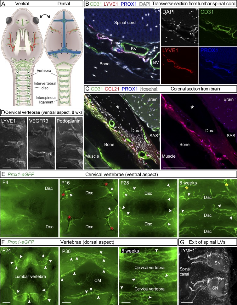
LVs in spinal meninges. (A) Schematic illustration of meningeal LVs (green) attached to the ventral and dorsal sides of the cranium and spinal canal after removal of the brain and spinal cord. SN; spinal nerve. (B) Transverse section of lumbar spine of an adult mouse showing LVs in dura, immunostained for CD31 (green), LYVE1 (red), and PROX1 (blue). (C) Coronal sections of adult skull showing the meningeal LVs, immunostained for CD31 (green), CCL21 (red), and PROX1 (blue). Asterisk indicates an artifactual space created during preparation. (D) LYVE1, VEGFR3, and podoplanin immunostaining of spinal meninges. (E and F) Development of the meningeal LVs in the spinal canal on the ventral (E) and dorsal (F) aspects during the indicated postnatal (P) days. (G) LVs exiting the spinal canal together with the spinal nerves. Arrowheads in E and F point to lymphatic valves, and red asterisks in E indicate BVs. Yellow asterisks in E indicate the connection between LVs around the FM and LVs around the spinal canal. Data shown are representative of n = 3–6 per time point and staining. Bars: (B) 20 µm; (C) 50 µm; (D, F, and G) 400 µm; (E) 300 µm.

