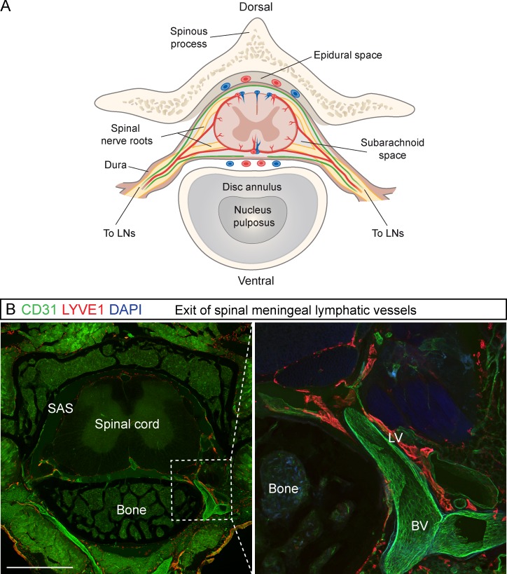
Figure 3.
LV exit from the spinal canal along the spinal nerves and BVs. (A) Schematic transverse view of the spinal cord and its blood (red) and lymphatic (green) vessels. (B) Transverse section of spinal cord with a close-up showing the exit of the LVs and BVs along the spinal nerve bundle in LYVE1 (red) and CD31 (green) immunostaining, respectively (n = 3). Bar, 2 mm.

