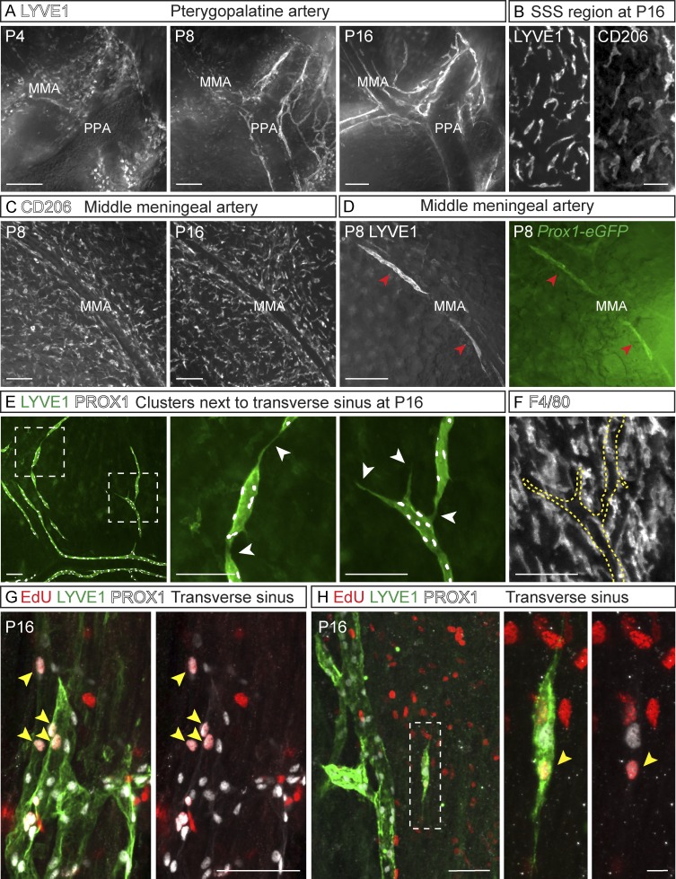
Figure 4.
Sprout extension and fusion of cell clusters in meningeal lymphangiogenesis. (A) LYVE1 staining of LVs developing around the PPA. (B) LYVE1+/CD206+ macrophage-like cells around the SSS at P16. (C) CD206 immunostaining around the MMA. (D) LYVE1 (gray) and Prox1-eGFP (green)–positive cell clusters (arrowheads) around the MMA. (E) LYVE1 (green) and PROX1 (gray) immunostaining of LEC clusters around the TS at P16. Dashed boxes indicate areas of the close-up images shown in E. Arrowheads indicate connections of the clusters with each other and with the already-formed LVs. (F) F4/80 immunostaining of macrophage-like cells for comparison. Dashed line indicates the LEC clusters shown in E. (G and H) EdU-positive LECs (arrowheads in G) in the tip cell area around the TS and (H) in isolated clusters stained for PROX1 (gray) and LYVE1 (green). EdU was administered 6 h before tissue harvest. Data shown are representative of n = 2–4 per time point and staining. Bars: (A and D) 200 µm; (B, F, and G) 50 µm; (C and E) 100 µm; (H) 10 µm.

