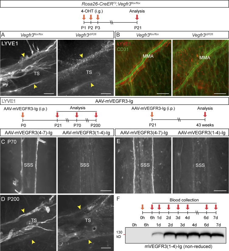
Figure 7.
VEGFR-3 is essential for meningeal LV development. (A and B) Comparison of dural LYVE1 staining in P21 mice deleted of Vegfr3 (Vegfr3iΔR26, n = 4) and their littermate controls (Vegfr3flox/flox, n = 9) around the TS (A) and MMA (B). BVs stained for CD31 (green). (C and D) LYVE1 staining around the SSS and TS in mice injected with the indicated AAVs at P0 and analyzed at P70 (n = 6, 6; C) or P200 (n = 3, 3; D). (E) LYVE1 staining around the SSS in mice injected with the same vectors on P21 and analyzed 40 wk later (n = 7, 6). The width of the TS is indicated by the arrowheads in A and D. (F) Western blot showing VEGFR3-Ig protein in serum of an individual mouse at the indicated time points after i.p. AAV injection. Data shown are representative of two independent experiments. Bars, 200 µm.

