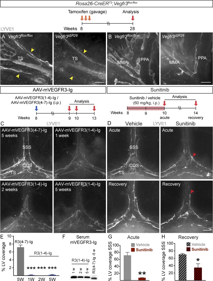
Figure 9.
VEGFR-3 signaling is required for LV maintenance in adult meninges. (A and B) Comparison of LYVE1 staining around the TS (A) and PPA (B) in Rosa26-Vegfr3flox/flox (n = 4) and Vegfr3iΔR26 mice (n = 4) 20 wk after tamoxifen administration. Arrowheads indicate TS width. (C and D) LYVE1 staining around the COS (C) at the indicated time points after AAV–mVEGFR31–4-Ig or AAV–mVEGFR34–7-Ig injection (n = 3, 3 in each time point) and in mice administered daily with 60 mg/kg sunitinib (D) and analyzed as indicated (n = 3, 3 in both time-points). Arrowheads point to the rostral end of the LV front in acute and recovery phases. (E) Quantification of the LV area in the experiment shown in C. (F) Western blot showing mVEGFR3-Ig protein in serum at the indicated time points after AAV injection. (G and H) Quantification of SSS length covered by LVs in sunitinib-treated mice in the acute phase (G) and recovery phase (H). Data shown are representative of two independent experiments. A Student’s t test was used to calculate p-values. *, P < 0.05; **, P < 0.01; ***, P < 0.001. Values are expressed as mean ± SEM. Bars: (A) 200 µm; (B) 150 µm; (C and D) 500 µm. W, weeks.

