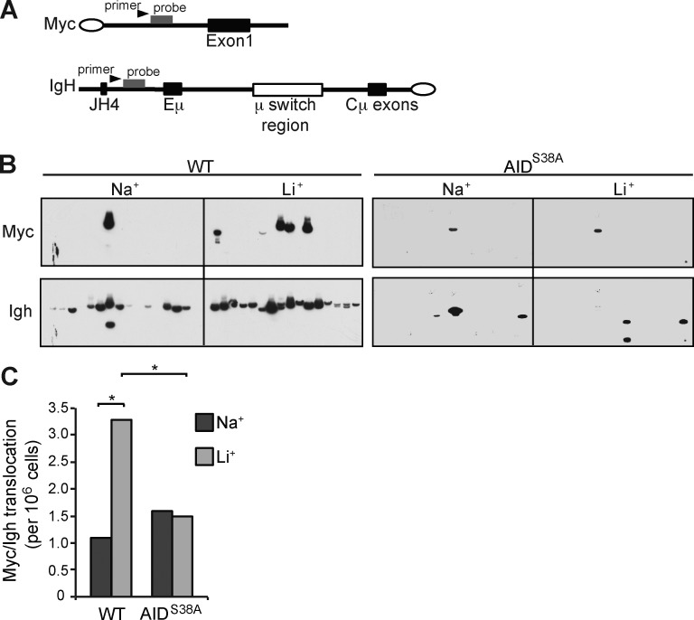
Figure 4.
AID Serine 38 phosphorylation promotes Myc/Igh translocations. (A) Schematic for the Myc/Igh translocation assay. PCR amplification primers are represented by black arrows and Southern probes by gray bars. Closed circles denote centromeric locations on the chromosomes. (B) Representative translocation assay Southern blots with Myc and Igh probes are displayed; each lane contains the DNA content of 1×10⁵ genomes. B cells from WT or AIDS38A mice cultured with IL-4 and LPS were treated with 10 mM NaCl or LiCl for 12 h before analysis. (C) Total translocation frequency summary from n = 4 independent experiments. P-values (*, P < 0.01) determined using two-tailed Fisher’s exact test (WT Na+ vs. AIDS38A Na+ or Li+, both P > 0.5).

