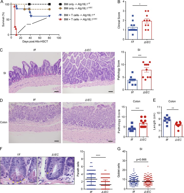
Figure 2.
Deletion of Atg16L1 in the intestinal epithelium of allo-HSCT recipients worsens GVHD. (A) Survival of lethally irradiated Atg16L1f/f (f/f) and Atg16L1ΔIEC (ΔIEC) mice transplanted with 5 × 10⁶ T cell–depleted BM cells with or without 2 × 10⁶ splenic T cells from donor B10.BR mice. n = 5 (f/f, BM only), 6 (ΔIEC, BM only), 13 (f/f, BM + T cells), and 15 (ΔIEC, BM + T cells). (B–E) Mice receiving BM and T cells from B10.BR mice as in A were sacrificed 4 d after allo-HSCT and analyzed for macroscopic signs of GVHD (B; see Materials and methods). Representative images of H&E-stained sections and pathology score of small intestine (C) and colon (D) and quantification of colon length (E). n = 10 mice per group. (F) Representative higher magnification H&E images from C and quantification of Paneth cells (yellow arrowheads). n = 5 mice per group. At least 50 crypts were quantified per mouse. (G) Quantification of goblet cells from sections in C stained with periodic acid–Schiff (PAS)/Alcian blue of the small intestine. At least 50 villi were quantified per mouse. n = 5 mice per group. (C, D, and F) Bars: 200 µm (C and D); 20 µm (F). Data points present individual mice in B–E, individual crypts in F, and individual villi in G. Bars represent mean ± SEM, and at least two independent experiments were performed. *, P < 0.05; **, P < 0.01; ***, P < 0.001; ****, P < 0.0001 by Mantel-Cox in A and Mann-Whitney U test in B–G.

