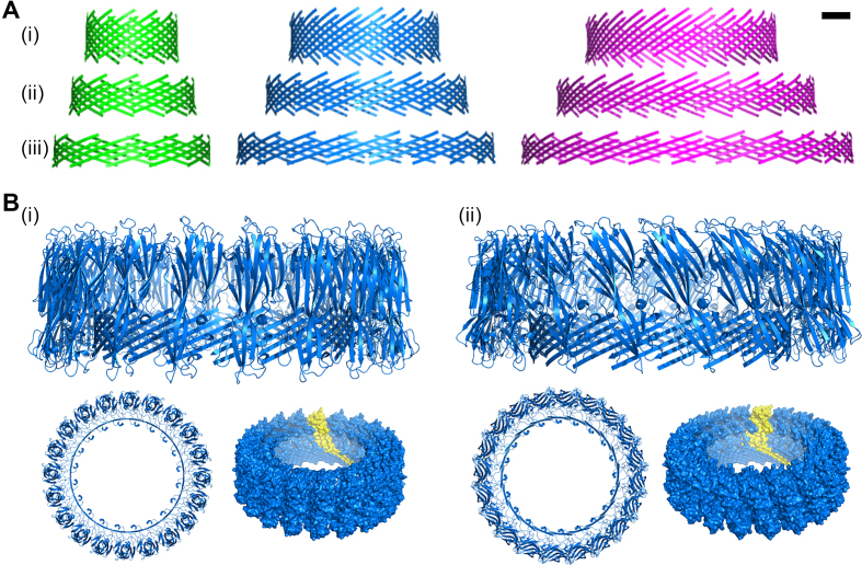
Figure 8.
Predicted structure of the DNA/αHL pore. (A) Modeled structure that represents the β-sheet forming residues of the transmembrane domain of a dodecameric (green), icosameric (blue) and hexacosameric (magenta) hybrid pore with an offset of (i) 4, (ii) 6, and (iii) 8 residues, respectively. Scale bar = 2 nm. (B) Possible structure formation of 20 αHL monomers with (i) an adjusted orientation of the protein cap domain to match the β-hairpin tilt for a 6-aa offset, and (ii) same alignment of the cap domain as for the natural occurring heptameric pore. Shown are the side view (top) and top view (bottom left) of the cartoon representation, and the surface representation with one monomer highlighted in yellow (bottom right).

