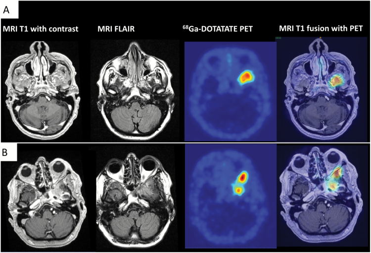
Fig. 3.
68Ga-DOTATATE PET for the differentiation of meningioma from tumor-free tissue and postoperative changes. (A) Preoperative imaging. MR and PET images of a 55-year-old male patient who presented with therapy-resistant left frontal headache for the past 6 months. T1-weighted MR images show subtle contrast enhancement at the skull base without exact delineation of tumor borders. The fluid attenuated inversion recovery sequence shows diffuse signal changes. In contrast, 68Ga-DOTATATE PET (DOTA-(Tyr3)-octreotide) is able to differentiate between meningioma and tumor-free tissue with an excellent tumor-to-background contrast. Fusion images were used for surgical planning. (B) Imaging at recurrence. Histological grading revealed a WHO grade II meningioma. After subtotal surgical resection (Simpson grade IV), the patient underwent radiation therapy. Follow-up imaging after 2 years revealed further tumor growth/recurrence. The T1-weighted image shows diffuse contrast enhancement at the skull base, difficult to be differentiated from postoperative changes. 68Ga-DOTATATE PET in contrast is consistent with meningioma tissue in this region with additional retrobulbar tumor growth.

