Figure 1.
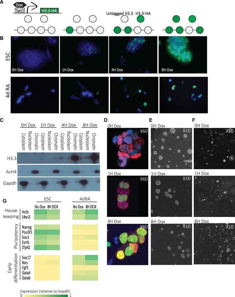
Genome-wide measurements of H3.3 dynamics. (A) A schematic diagram showing components of the doxycycline (Dox) histone H3.3-HA induction system in KH2 ESC and the use of this system for assaying H3.3 turnover dynamics. (B) HA-tagged H3.3 expression in ESC (upper panel) and in 4d RA cells (lower panel) was induced by Dox addition at different times and immunolabeled with anti-HA antibodies. DAPI staining overlay is shown in blue. (C) Time course western blot following fractionation of ESC cellular extracts to three fractions showing protein levels of transgenic HA-H3.3 (lower panel) compared to acetylated H4 (upper panel, chromatin bound) and GAPDH (middle panel, cytoplasmatic) expression at time points from 0 to 8 h (0–8 h) after Dox addition. (D) HA (green) and Bromodeoxyuridine (BrdU) (red) immunostaining of KH2 ESCs treated with IdU for 2 h and Dox for 0, 1 and 8 h. Nuclei were stained with DAPI and analyzed by confocal microscopy. (E) Representative bright-field images of KH2 ESCs after 0, 1 and 8 h with Dox show no significant difference in colonies morphology. (F) Representative bright-field images of 4dRA cells after 0, 1 and 8 h with Dox show no significant difference in cell morphology. (G) Expression levels of pluripotency, selected lineage marker and housekeeping genes in KH2 ESCs and 4dRA cells, with and without Dox addition. Normalized to Gapdh expression.

