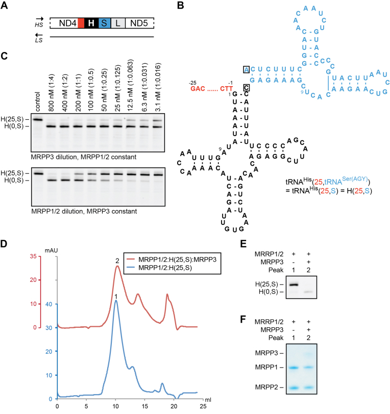
Figure 2.
Role of MRPP1/2 in RNase P function on tRNAHis. (A) Schematic representation of the heavy-strand (HS) tRNA cluster His-Ser(AGY)-Leu(CUN). The 5′- and 3′-region of tRNAHis (in black) are highlighted in red and blue, respectively. (B) Secondary structure of pre-tRNAHis(25,tRNASer(AGY)) (H(25,S)) having 25 nt as 5′-leader (in red) and the complete tRNASer(AGY) as 3′-trailer (in blue). The discriminator bases are boxed. (C) RNase P dilution series on 200 nM tRNAHis(25,S). Top: MRPP3 dilution (from 800 to 3.1 nM) and MRPP1/2 constant (800 nM). Bottom: MRPP1/2 dilution (from 800 to 3.1 nM) and MRPP3 constant (50 nM). The molar ratio of tRNA to protein is shown in parentheses. The reactions were incubated for 1 h. (D–F) Gel filtration chromatograms of MRPP1/2:pre-tRNAHis(25,S) prior and after MRPP3 treatment (D). Peaks of interest are numbered, and tRNA and protein content of the peaks are visualized by urea PAGE (E) and SDS-PAGE (F), respectively.

