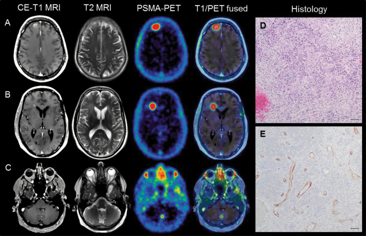
Fig. 1.
(A–C) Examples of the high uptake in PSMA PET; even a lesion in the cerebellum with a small diameter of 0.3 cm in MRI could be visualized by PSMA PET due to its extraordinarily high tumor-to-background ratio (TBR) contrast. (D) Gliosarcoma (WHO grade IV) showing high cellularity, pleomorphism, and microvascular proliferation. (E) Immunohistochemical PSMA staining revealed a strong PSMA expression throughout the tissue specimen, specifically in neovascular endothelial cells without any expression in solid tumor tissue (scale bar: 50 µm).

