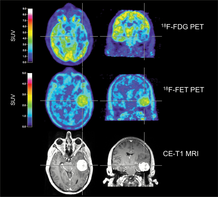
Fig. 1.
A 43-year-old male patient with a newly diagnosed left temporal meningioma (WHO grade I), preoperatively examined by multimodal imaging. Both contrast-enhanced MRI and 18F-FET PET allow a precise tumor delineation. Conversely, 18F-FDG PET shows decreased metabolic activity, indicating its limitation for the evaluation of meningioma extent.

