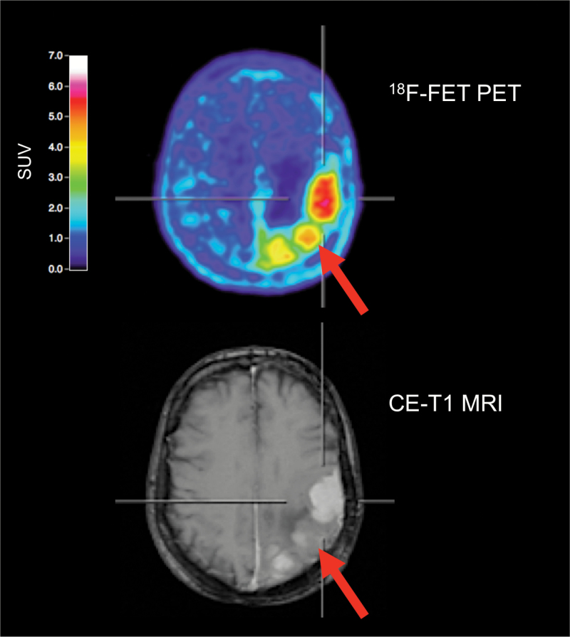
Fig. 4.
Amino acid PET with 18F-FET and contrast-enhanced MR images of a 68-year-old female meningioma patient (WHO grade I) with suspected recurrence 9 years after tumor resection at initial diagnosis. 18F-FET PET identifies 3 hypermetabolic lesions, consistent with meningioma recurrence. In contrast, MRI shows prominent contrast enhancement in only 2 of 3 lesions. In that lesion (arrow, bottom), contrast enhancement is subtle and not well defined. 18F-FET PET allows an improved delineation of this lesion (arrow, top).

