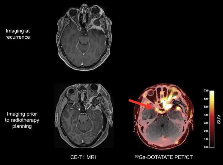
Fig. 5.
A 42-year-old patient with exophthalmos and a history of a left sphenoid wing meningioma. Preoperative MRI shows tumor recurrence (top). Postoperative MRI (bottom left) shows an incomplete resection of the tumor, necessitating adjuvant radiotherapy. For radiotherapy planning, 68Ga-DOTATATE PET/CT reveals an additional tumor located at the tip of the right sphenoid wing (arrow, bottom right).

