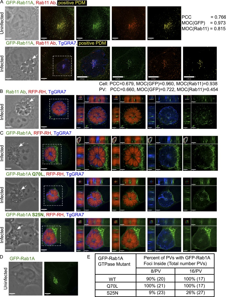
Figure 1.
Analysis of host Rab11 and Rab1A foci in the PV. (A) VERO cells expressing GFP-Rab11A (green) were infected with parasites for 24 h and immunostained for Rab11 (red) and TgGRA7 (blue). To measure the level of colocalization, a positive product of the difference of the means (PDM; yellow) was calculated for uninfected and infected cells with PCC and MOC for the GFP and Rab11 channels. (B) VERO cells infected with RFP expressing parasites for 24 h (RFP-RH; red) were immunostained for Rab11 (green) and TgGRA7 (blue). (C) HeLa cells expressing GFP-Rab1A, the GTP-locked mutant Q70L, or the GDP-locked mutant S25N (green) were infected with RFP-RH for 24 h (16 parasites/PV) and immunostained for TgGRA7 (blue). (D) An uninfected, GFP-Rab1A expressing HeLa cell shows Rab1A in the ER–Golgi. (E) Percentage of PVs containing intra-PV GFP-Rab1A foci for one representative experiment. For all images, individual z-slices are shown; the boxed region is magnified in the orthogonal views, highlighting the PV (arrows). Bars: 6 μm; (orthogonal views) 1.7 μm.

