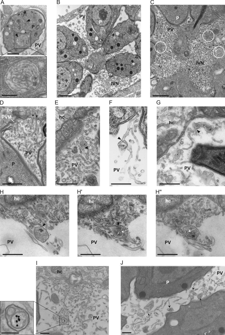
Figure 4.
Localization of host LDL–containing endosomes and the IVN. (A–D) TEM images of infected VERO cells. (A) Intracellular compartments containing membrane tubules likely of the IVN before secretion (arrowheads). Bars: 200 nm; (inset) 100 nm. (B) Intravacuolar distribution and morphology of the IVN tubules after secretion. Bar, 500 nm. (C) IVN at the center of a PV with membrane-bound structures containing vesicles likely originating from the host cell (white circles). Bar, 150 nm. (D) Connection of IVN tubules with the PVM (arrows). Bar, 200 nm. (E–J) TEM images of infected VERO cells incubated with LDL-gold for 24 h. (E–G) Detection of intra-PV host LDL-containing endocytic structures within a tubule of the IVN (arrowheads). Bars, 200 nm. (H–H′′) Shown are serial sections of an intra-PV, membrane-bound structure that is attached to an IVN tubule and contains several host LDL–containing vesicles (arrows). Bars, 150 nm. (I and J). Membrane-bound structures, unattached to IVN tubules, with host LDL vesicles (arrows) in regions enriched in the IVN. Bars, 200 nm. hc, host cell; P, parasite.

