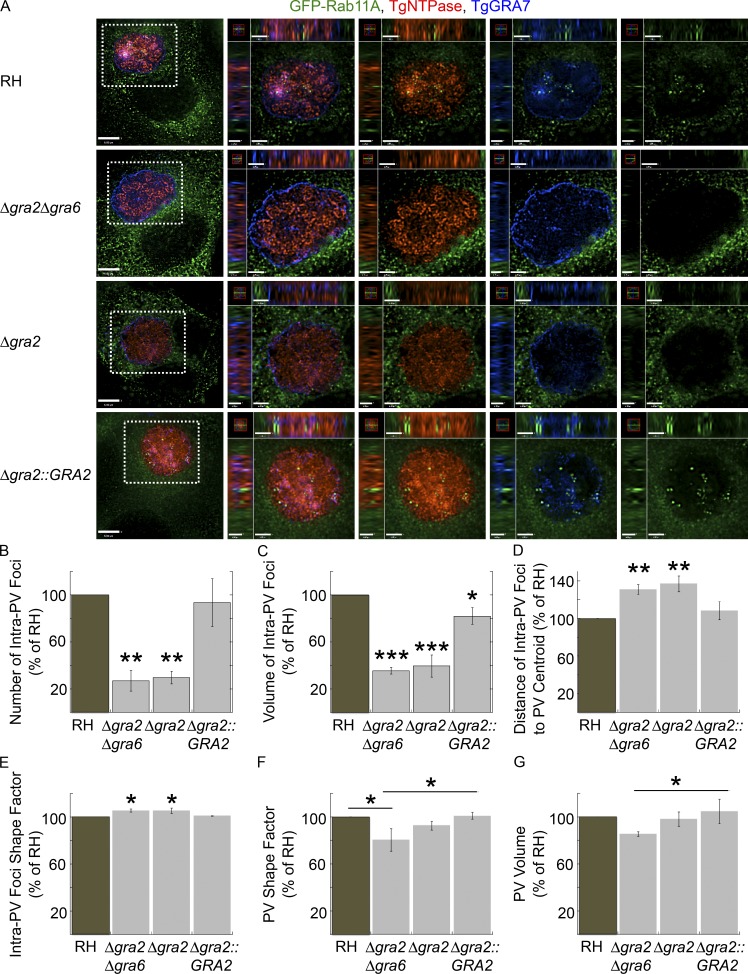
Figure 5.
Role of TgGRA2 and TgGRA6 on the internalization of host GFP-Rab11A into the PV. (A–D) VERO cells expressing GFP-Rab11A (green) were infected with RH, Δgra2Δgra6, Δgra2, and Δgra2::GRA2 parasites for 24 h and then fixed and immunostained for TgNTPase (red) and TgGRA7 (blue). (A) Individual z-slices are shown with the boxed region magnified in the orthogonal views, highlighting the PV. Bars: 6 μm; (orthogonal views for RH and Δgra2Δgra6) 1.3 μm; (orthogonal views for Δgra2 and Δgra2::GRA2) 1.4 μm. (B–G) The number (B), volume (C), distance from the PV centroid (D), and shape factor (E) of intravacuolar GFP-Rab11A foci with the shape factor (F) and volume (G) of PVs calculated for PVs containing 16 parasites. Mean values ± SD, n = 3 independent experiments. PVs measured: RH (23–25), Δgra2Δgra6 (19–22), Δgra2 (19–21), and Δgra2::GRA2 (23–26). *, P < 0.03; **, P < 0.001; ***, P < 0.0001.

