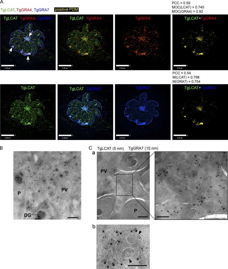
Figure 7.
Localization of the Toxoplasma lipolytic lecithin/cholesterol acyltransferase (TgLCAT) to the IVN. (A) Infected HFF cells (24 h) were immunostained for TgLCAT (green), TgGRA4 (red), and TgGRA7 (blue), and a positive product of the difference of the mean (PDM) image (yellow), PCC, and MOC were calculated. Arrows indicate TgLCAT colocalized with the IVN. Bars, 6 μm. (B and C) Immuno-EM of HFF cells infected with Δlcat:LCAT-HA with anti-HA for 24 h showing TgLCAT on the IVN colocalized with TgGRA7 (a) and on double membrane structures (b). Arrowheads in b point to internal vesicles likely from the host cell surrounded by the PVM. Bars: 200 nm; (a, inset) 100 nm.

