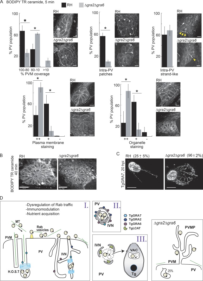
Figure 9.
Distribution of host derived ceramides in the PV and parasites. (A and B) HFF cells infected with RH or Δgra2Δgra6 parasites (20 h) were incubated with BODIPY TR C5-ceramide for 5 min (A; bars, 3 µm) or 40 min (B; bars, 7 µm) and fixed and viewed by fluorescence microscopy. Individual z-slices are shown. For the 5-min treatment, PVs containing eight parasites were placed into categories based on their staining pattern as described in Materials and methods. Arrowheads in A show filamentous structures connecting the PVM with parasites. *, P < 0.005. (C) HeLa cells were infected with RH or Δgra2Δgra6 parasites (20 h) and immunostained for TgGRA7 to detect PVM projections (PVMPs). Total intensity projections of z-stacks and percentage of the PV population with PVMPs (mean ± SD) are shown. *, P < 0.0001. Bars, 7 µm. (D) Hypothetical model of host vesicles internalized into the PV in WT (left cartoons) and Δgra2Δgra6 parasites. H.O.S.T., host organelle–sequestering tubulostructures.

