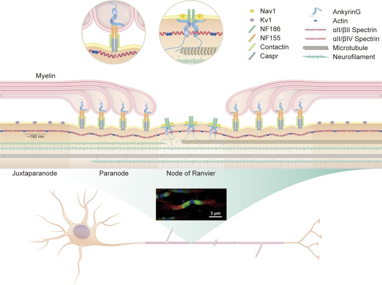
Figure 1.
Architecture of axonal cytoskeleton and polarized molecular domains. Myelination induces architectural rearrangement of the axon into polarized molecular domains. The nodes of Ranvier are short, unmyelinated segments that contain clusters of voltage-gated sodium channels. Flanking the nodes are the paranodes, junctions between noncompacted paranodal myelin loops and the underlying axolemma. Distal to the paranodes are the juxtaparanodes, which contain voltage-gated potassium channels. The fluorescent micrograph of optic nerve axons illustrates the distinct localization of potassium channels (red, juxtaparanode), Caspr (green, paranode), and βIV spectrin (blue, node). The axon cytoskeleton facilitates structural integrity and molecular organization and acts as a conduit for axon transport. It consists primarily of neurofilaments, microtubules, actins, and spectrins. Actins and spectrins assemble into a repeating lattice with structural periodicity of 180 to 190 nm. At nodes, ankyrin adaptor proteins anchor nodal constituents such as voltage-gated ion channels to the underlying actin–spectrin cytoskeleton. At paranodes, protein 4.1B (not depicted), anchors the NF155–Caspr–Contactin complex to the underlying actin–spectrin cytoskeleton.

