Figure 6.
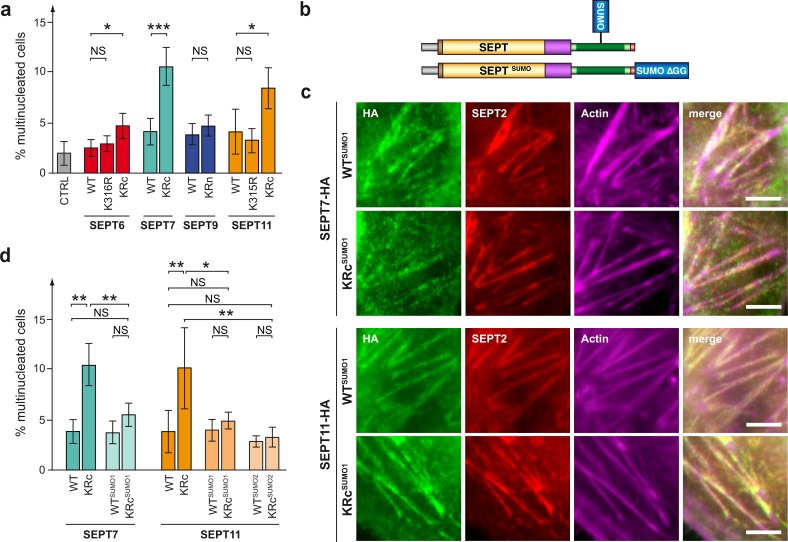
Role of septin SUMOylation in cell division. (a) Percentage of HeLa cells exhibiting multinucleation after transfection with a control plasmid (pCDNA.3; CTRL) or expression vectors for WT or mutant septins (mean from three to five independent experiments). (b) Schematic representation of SUMOylated HA-tagged septin (top) or septin C-terminally fused to SUMO (bottom). (c) Fluorescent light microscopy images of septin filaments formed by WT or non-SUMOylatable SEPT7 and SEPT11 mutants C-terminally fused to SUMO in HeLa cells. Cells were stained for HA-tagged septins (anti-HA antibodies, green), endogenous SEPT2 (anti-SEPT2 antibodies, red), and actin (phalloidin, magenta). Bars, 5 µm. (d) Percentage of HeLa cells exhibiting multinucleation after transfection with WT, non-SUMOylatable, or constitutively SUMOylated SEPT7 and SEPT11 (mean from three to six independent experiments). Error bars, SD; *, P < 0.05; **, P < 0.01; ***, P < 0.001.

