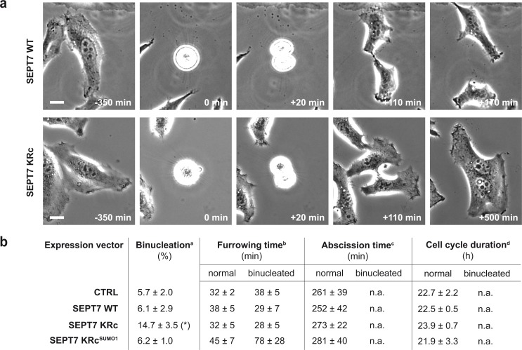
Figure 7.
Lack of septin SUMOylation interferes with late steps of cytokinesis. (a) Representative images from time-lapse microscopy analysis of HeLa cells expressing WT or non-SUMOylatable SEPT7. Images correspond to different cell division events including cell rounding, cleavage furrow ingression, intercellular bridge formation, and either bridge abscission (top) or bridge regression and formation of a binucleated cell (bottom). Bars, 10 µm. (b) Time-lapse microscopy analysis of HeLa cells transfected with control plasmid (pCDNA.3; CTRL) or expression vectors for SEPT7 WT, KRc, or KRcSUMO1 variants. 24 h after transfection, time-lapse sequences of cells were recorded every 20 min for 48 h. At least 50 transfected cells were analyzed for each condition in each experiment. Means ± standard errors from three independent experiments are indicated. *, P < 0.05 compared with control condition (two-tailed two-sample equal-variance Student’s t test). aPercentage of cells for which cell division results in the formation of binucleated cells. bTime between cell rounding and furrow ingression. cTime between cell rounding and intercellular bridge abscission. dTime between two cell-rounding events. n.a., not applicable.

