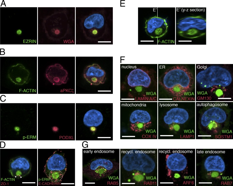
Figure 1.
The apicosome is a unique cellular structure. (A–D) Fluorescent confocal images of single H9 cells stained with indicated markers. (E) Fluorescent confocal images of single H9 cells stained for phalloidin (green, F-ACTIN). Image in E′ is the optical y-z section (indicated by white lines) of the 3D rendered image of the cell in E. (F and G) Singly isolated H9 cells are stained for membrane (green, WGA) as well as antibodies specific to distinct organelles (red) as indicated. Bar, 10 µm. For all images, blue indicates DNA staining (HOECHST).

