Figure 4.
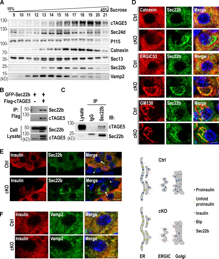
cTAGE5 interacts with v-SNARE Sec22b and controls its localization. (A) Subcellular fractionation of MIN6 cells using a continuous sucrose gradient. Data were from three independent experiments with similar results. (B) The interaction of Sec22b with cTAGE5 was assessed by coIP. Constructs expressing Sec22b and cTAGE5 were transfected alone or in combination into 293 cells; 24 h later, cell lysates were precipitated with Flag agarose beads and the immune complexes were detected with Flag or GFP antibody. (C) The interaction between endogenous Sec22b and cTAGE5 in pancreatic islets was observed by coIP with Sec22b antibody. (D) Pancreas sections immunostained with Sec22b (green), Calnexin (red), ERGIC53 (red), and GM130 (red). Data were from three independent experiments with similar results. Bar, 10 µm. The model of distribution of Sec22b throughout ER-to-Golgi transport pathway shown under the figure. (E) Pancreas sections stained with insulin (red) and Sec22b (green). The arrow shows β cell in islets; the arrowhead shows non–β cells in islets. Bar, 20 µm. (F) Pancreas sections stained with insulin (red) and Vamp2 (green). Bar, 10 µm.

