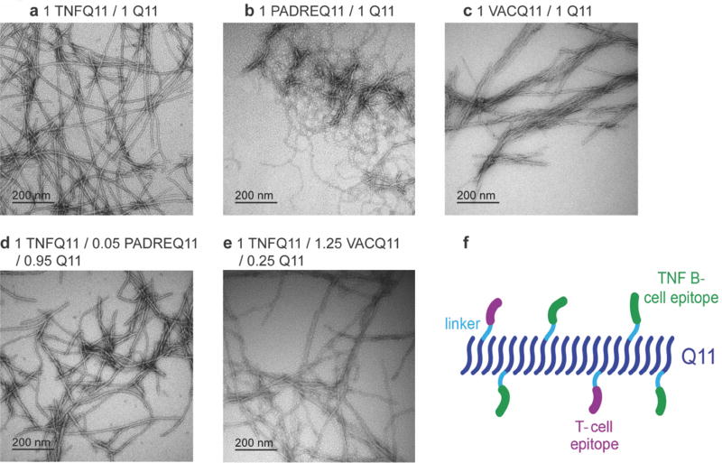
Fig 1. TEM and schematic of co-assembled peptide nanofibers.
Negative-stained TEM images of nanofibers (prepared at 2 mM total peptide concentration and diluted to 0.2 mM for imaging) (a) TNFQ11/Q11 (1:1 molar ratio), (b) PADREQ11/Q11 (1:1), (c) VACQ11/Q11 (1:1), (d) TNFQ11/PADREQ11/Q11 (1:0.05:0.95), (e) TNFQ11/VACQ11/Q11 (1:1.25:0.25). (f) schematic of co-assembled peptide nanofibers containing B-cell epitopes and T-cell epitopes.

