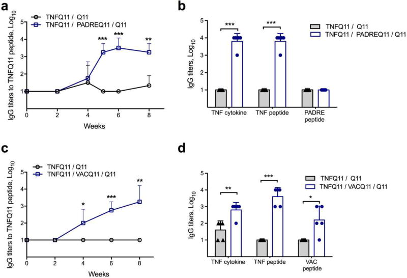
Fig 2. Co-assembled peptide nanofibers raise anti-TNF antibodies.
(a) TNFQ11 peptide-specific IgG responses in the sera of mice immunized with formulations with or without the self-assembling T-cell epitope PADREQ11, evaluated over time by ELISA. (b) Reactivity of immune sera against TNF cytokine (mouse), TNF4–23 peptide, and PADRE peptide. (c) TNFQ11 peptide-specific IgG responses in the sera of mice immunized with formulations with or without the self-assembling T-cell epitope VACQ11, evaluated over time by ELISA. (d) Reactivity of immune sera against TNF cytokine, TNF4–23 peptide, and VAC peptide. Only T-cell epitope-containing assemblies raised significant IgG responses against TNF peptide and TNF cytokine, and there was minimal antibody development against the T-cell epitopes PADRE or VAC. Formulations were 1 mM TNFQ11/0.05 mM PADREQ11/ 0.95 mM Q11 (a–b), 1mM TNFQ11/0.25 mM VACQ11/0.75 mM VACQ11 (c), and 1mM TNFQ11/1.25 mM VACQ11/0.25 mM Q11 (d). In (a, c) mice were immunized at week 0 and boosted at week 4 with a half dose. In (b, d), titers were measured after priming and two boosts at weeks 4 and 8. Mean ± SD is shown. n=4 for each group in a and c. n=5 for each group in b and d. Statistical significance was confirmed by t-test at each time point (a and c) or for each antigen (b and d). * p<0.05, ** p<0.01, *** p<0.001.

