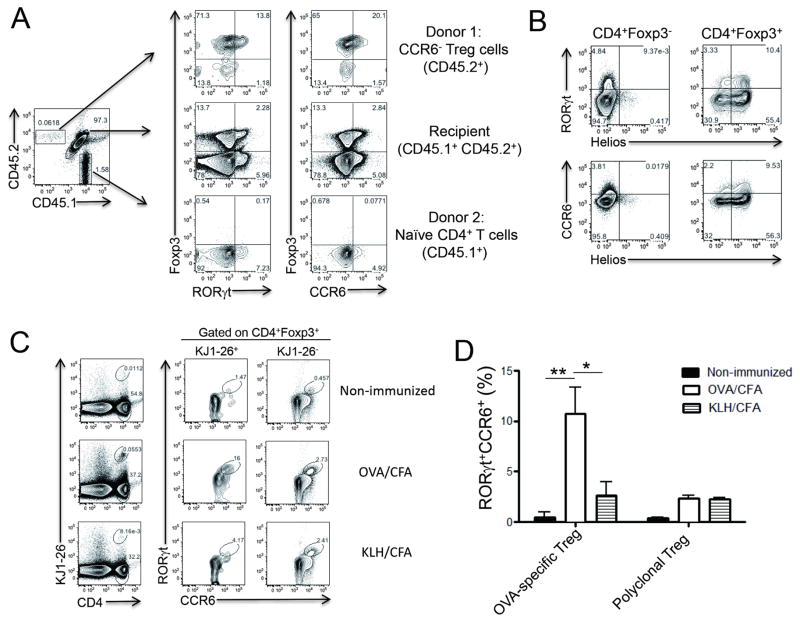
Figure 3. Origin of RORγt+CCR6+ Treg cells.
(A) CD4+YFP+CCR6− Treg cells isolated from Foxp3YFP-Cre reporter mice (CD45.2+) were mixed with CD4+CD25−CD44loCD62L+ naïve T cells isolated from B6.SJL mice (CD45.1+) in the 1:9 ratio and then were adoptively transferred to CD45.1+CD45.2+ recipient mice. One day after the transfer, recipient mice were subcutaneously immunized with MOG/CFA. Seven days later, CD4+ T cells in dLNs were analyzed for Foxp3, RORγt and CCR6 expressions after gating populations based on CD45.1 and CD45.2 expression. (B) Helios expression in CD4+Foxp3+ or CD4+Foxp3− cells in dLNs was compared with RORγt or CCR6 expression. (C–D) OVA-specific CD4+KJ1-26+GFP+CCR6− Treg cells isolated from DO11.10xFoxp3GFP reporter mice were adoptively transferred to BALB/c mice. One day after the transfer, mice were s.c. immunized with OVA/CFA or KLH/CFA or left untreated. Seven days after immunization, KJ1-26+ or KJ1-26− CD4+Foxp3+ Treg cells in dLNs were analyzed for RORγt and CCR6 expressions. Data are representatives of two independent experiments. *, p<0.05. **, p<0.005. See also Figure S2.

