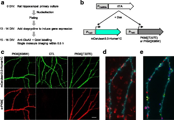
Fig. 1.

Overexpression of active or inactive PKMζ in cultured rat hippocampal neurons and single molecule imaging of GluA2-containing AMPARs. a Experimental workflow. b Transgene expression using TetON system. c Representative image of fluorescence microscopy of Homer1C-fused mCerulean3.0 and anti-protein kinase C, zeta (PKCζ) immunofluorescence signals in neurons expressing active PKMζ (T227E), inactive PKMζ (K98W), or neither (CTL). d Representative image of Qdot-labelled GluA2-containing AMPARs (red, maximum intensity projection of 500 frames) and mCerulean3.0 (cyan, average image of 100 frames). (e) Qdot trajectories (open circles) observed in the dendritic region (cyan)
