Figure 5.
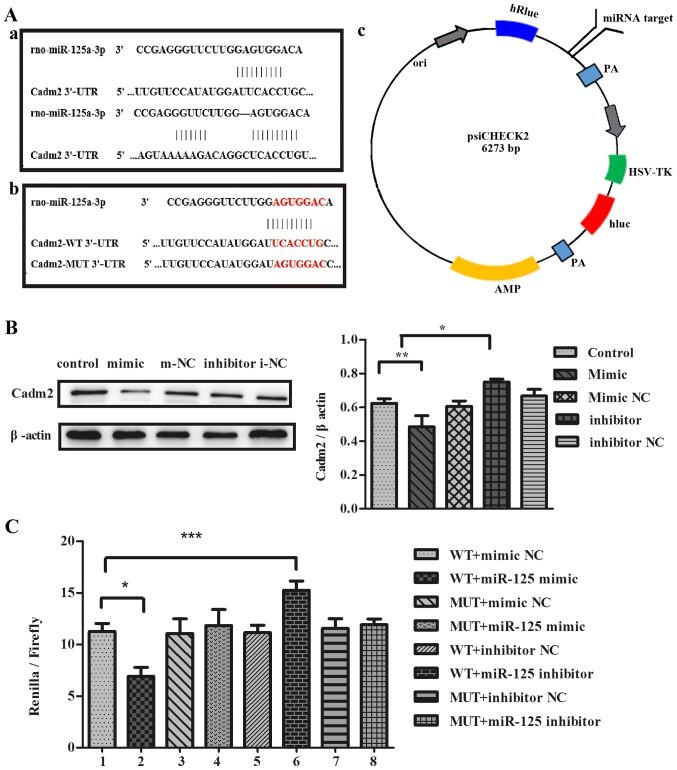
miR-125a regulates the expression of Cadm2. (A) (a) The target sites of miR-125a in the Cadm2 3′-UTR were predicted by bioinformatics software, TargetScan and miRNA.org. (b) The target sites of miR-125a in Cadm2-WT 3′-UTR and Cadm2-MUT 3′-UTR. (c) The construction profile of the psiCHECK2-Cadm2 vector is presented in the diagram that contained the miR-125a-3p target sites in Cadm2 3′-UTR. (B) Expression of target protein Cadm2 in the PC12 cells were analyzed with the western blot assay, and the bands were quantified with ImageJ software. One-way analysis of variance was used to analyze the variances. By western blotting, the expression of the Cadm2 protein was efficiently decreased by miR-125a mimics, compared with the control group, and may be increased obviously when treated with miR-125a inhibitor. (C) The miR-125a mimic, inhibitor, mimic NC or inhibitor NC were co-transfected with Cadm2-WT 3′-UTR or Cadm2-MUT 3′-UTR into PC12 cells. miR-125a targeted the expression of Cadm2. One-way analysis of variance test was used to analyze the variances between groups. *P<0.05, **P<0.01 and ***P<0.001 as indicated. miR, microRNA; UTR, untranslated region; WT, wild-type; NC, negative control; Cadm2, cell adhesion molecule 2.

