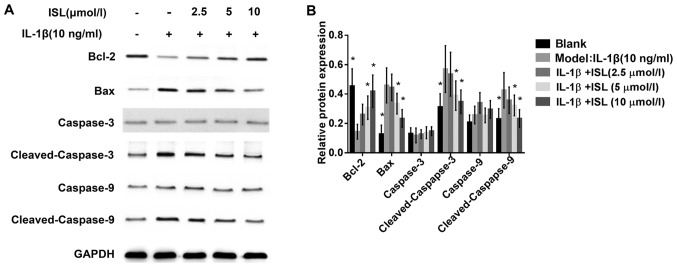
Figure 6.
The effect of ISL on the levels of anti-apoptotic and pro-apoptotic proteins in IL-1β-induced chondrocyte-like ATDC5 cells. (A) The IL-1β stimulated the cells were treated with different doses of ISL (2.5, 5 and 10 µmol/l) for 48 h. Then protein expressions Level of Bcl-2, Bax, caspase-3, cleaved-caspase-3, caspase-9 and cleaved-caspase-9 were detected by western blotting. (B) The protein analysis of quantitative fluorescence. GAPDH was used as internal control. The values represent the mean ± standard deviation of three independent experiments. *P<0.05 vs. model group treated with only IL-1β. ISL, isoliquiritigenin; IL, interleukin.

