
Fig. 3. T cell clones differ in their response against M. tuberculosis MA forms.
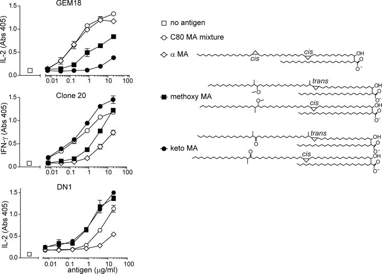
GEM18, clone 20, and DN1 T cells (5×10⁴) were stimulated with allogeneic monocyte-derived dendritic cells (5×10⁴) and antigen in duplicate wells. After 20 hours, supernatants were tested for the presence of IL-2 (GEM18) or IFN-γ (clone 20 and DN1). Structures of α-, keto-, and methoxy MA are drawn as previously reported and represent the average of a naturally occuring range of tail lengths [24, 34]. One out of two independent experiments is shown. Cytokine standard curves are shown in Supplemental Fig. 5. Error bars indicate SEM.