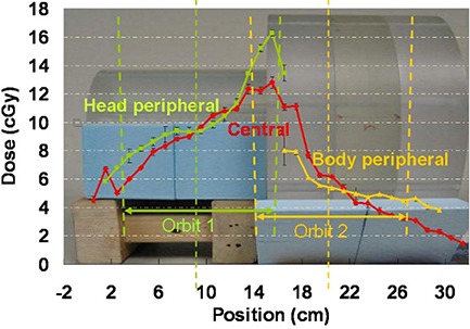
Figure 8.

Central channel and peripheral channel dose profiles overlaid with the imaging geometry. The figure shows the dose measured using the OBI 1.3 standard dose scans. For the OBI 1.3 low‐dose scans and the OBI 1.4 low‐dose thorax scans that we have selected for our head and neck protocol patients, the dose should be scaled to about 1/5 and 1/10, respectively.
