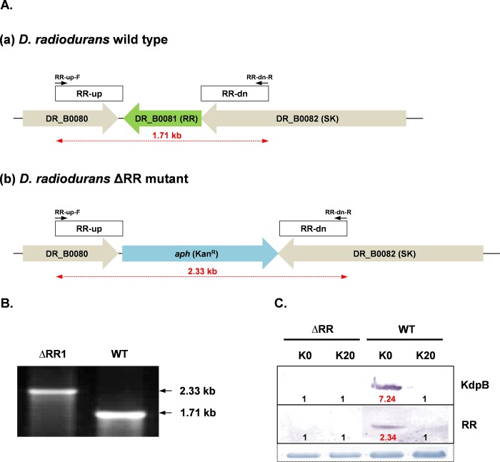
Fig 3. Construction and confirmation of ΔRR mutant.
(A) Schematic representation of the RR gene (DR_B0081) in wild type D. radiodurans (a) and its replacement with kanamycin resistance cassette (aph) in ΔRR D. radiodurans (b). The primers used for the PCR confirmation of the mutant are shown. (B) Confirmation of complete deletion of RR gene in ΔRR D. radiodurans as compared to wild type D. radiodurans, using primer pair shown in Fig 3A. (C) Expression of KdpB or RR proteins in wild type or ΔRR D. radiodurans cells incubated in K0 or K20 media. Details of immuno-staining, loading controls and fold change levels were same as described in legend to Fig 2.

