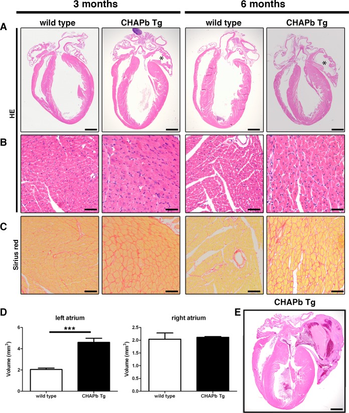
Fig 2. Hypertrophy and left atrial enlargement in CHAP Tg hearts.
(A-C) Wt (left panels) and CHAPb Tg (right panels) at 3 and 6 months of age. (A) HE stained overview section. The left atrium in CHAPb Tg hearts is enlarged (indicated by *) compared to wt litter mates. (B) Higher magnification of left ventricle. In the left ventricle of CHAPb Tg hearts the cardiomyocytes are hypertrophic. (C) Sirius red staining of the left ventricle showing increase in interstitial fibrosis in CHAPb Tg. (D) Myocardial volume of the left atrium (left panels) and right atrium (right panels) in wt (white bars) and CHAPb Tg (black bars) hearts at 3 months of age (wt n = 4, Tg n = 4, T-test p< 0.001). (E) CHAPb Tg heart with severe phenotype showing pronounced atrial enlargement, filled by a thrombus and thickening of the ventricles. Scale bars 1 mm in A and D, 50 μm in B, C.

