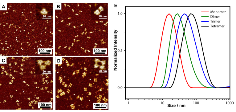
Figure 4.
AFM and DLS analysis of the tetrahedron assemblies. (A–D) The fluid mode AFM images of the tetrahedron monomer (A), dimer (B), trimer (C) and tetramer (D). The inset is the amplified single structure. (E) The DLS profile of the tetrahedron monomer (red), dimer (green), trimer (blue) and tetramer (black).

