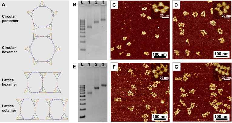
Figure 5.
AFM and 2.5% agarose gel electrophoresis of the circular pentamer, hexamer and the lattice hexamer and octamer. (A) The schematic illustration of the larger circular structures. (B) 2.5% agarose gel electrophoresis of circular pentamer and hexamer. Lane L: DNA ladder with 100–1000 base pairs. Lane 1: circular tetramer as a control; Lane 2 and 3: circular pentamer and hexamer respectively. (C and D) The fluid mode AFM images of circular pentamer and hexamer. The inset is the amplified single structure. (E) 2.5% agarose gel electrophoresis of lattice hexamer and octamer. Lane L: DNA ladder with 100–1000 base pairs. Lane 1: circular tetramer as a control; Lane 2 and 3: lattice hexamer and octamer respectively. (F and G) The fluid mode AFM images of lattice hexamer and octamer. The inset is the amplified single structure.

