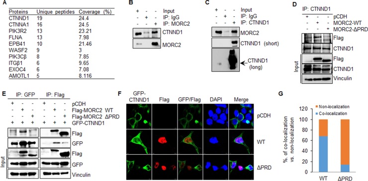
Figure 5. MORC2 interacts with CTNND1 through its PRD domain.
(A) The proteins that specifically interacted with Flag-MORC2 WT were analyzed by matching unique peptide numbers and the percentage of coverage. (B, C) HEK293T cells were subjected to the sequential IP-Western blot analysis with the indicated antibodies. (D) HEK293T cells stably expressing pCDH, Flag-MORC2 WT, and Flag-MORC2 ΔPRD were subjected to IP analysis with an anti-CTNND1 antibody, followed by immunoblotting with the indicated antibodies. (E) HEK293T cells were transfected with the indicated expression vectors. After 48 h of transfection, total cellular lysates were subjected to sequential IP-Western blot analysis with the indicated antibodies. (F, G) HEK293T cells were co-transfected with the plasmids encoding pCDH, Flag-MORC2 WT, and Flag-MORC2 ΔPRD along with GFP-CTNND1, and then immunofluorescence staining was carried out using an anti-Flag antibody (F). Cell nuclei were counterstained with DAPI. The quantitative results of co-localization of GFP-CTNND1 with Flag-MORC2 and Flag-MORC2 ΔPRD are shown in G.

