
Figure 2. Localization of CSF-1R and TSC-22.
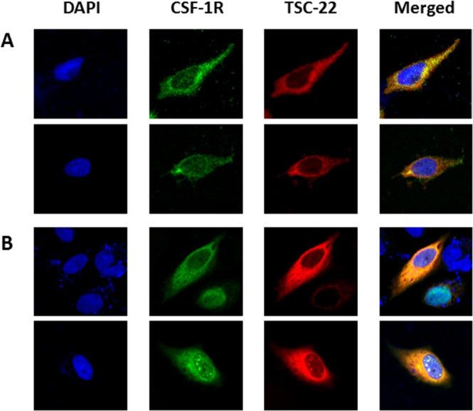
HeLa cells were non-transfected (A) or co-transfected with pcDNA4-TSC-22 and CSF-1R (B). ICC was performed using CSF-1R (green) and TSC-22 (red) antibodies, and their co-localization was represented by a yellow color. Cell images were taken using a confocal fluorescence microscope (magnification ×400).