
Figure 3. Protein levels of CSF-1R and TSC-22 in cervical cancer cells.
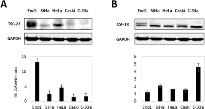
(A) Total protein was prepared from each indicated cell line. The expression of TSC-22 protein was analyzed using western blotting. Expression levels of TSC-22 were normalized to GAPDH expression. (B) Whole-cell lysates were prepared from the same cell line indicated in A and used in western blot analysis. CSF-1R expression levels are presented as the CSF-1R/GAPDH ratio and compared with a normal cervical cell line (End1).